In the realm of medical imaging, advancements continue to revolutionize our ability to visualize and understand the intricate structures of the human body. Among these innovations, Magnetic Resonance Angiography (MRA) stands out as a powerful tool for non-invasively imaging blood vessels with exceptional detail and clarity. By harnessing the principles of magnetic resonance imaging (MRI), MRA provides invaluable insights into vascular anatomy, aiding in the diagnosis and treatment of a wide range of cardiovascular conditions. In this comprehensive exploration, we delve into the mechanics, applications, and advancements of Magnetic Resonance Angiography, shedding light on its profound impact in modern medicine.
Understanding Magnetic Resonance Angiography:
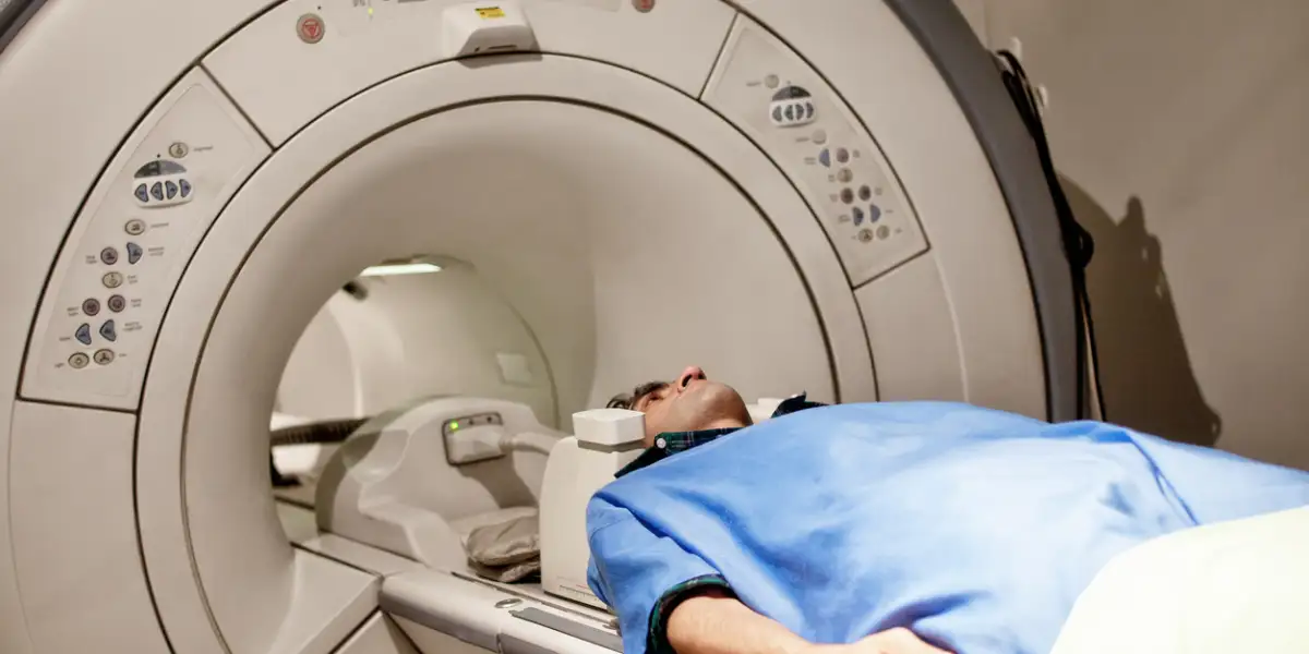
Magnetic Resonance Angiography (MRA) is a non-invasive imaging technique that utilizes magnetic resonance imaging (MRI) technology to visualize blood vessels throughout the body. Unlike conventional angiography, which requires the injection of contrast agents and invasive catheterization, MRA relies on the intrinsic properties of blood and magnetic fields to generate detailed images of vascular structures. By selectively enhancing the signal from flowing blood, MRA allows for the visualization of arteries and veins with high spatial resolution and soft tissue contrast.
Techniques and Modalities:
Magnetic Resonance Angiography encompasses various techniques and modalities, each offering unique advantages and applications:
- Time-of-Flight (TOF) MRA: This technique relies on the inflow of unsaturated blood into the imaging plane to create contrast between flowing blood and stationary tissue. By selectively saturating stationary tissue signals, TOF-MRA accentuates the signal from flowing blood, resulting in bright vessel images with excellent spatial resolution.
- Phase-Contrast MRA: Phase-contrast MRA measures the phase shift of moving blood relative to stationary tissue, allowing for the quantification of blood flow velocity and direction. This technique is particularly useful for evaluating blood flow dynamics and detecting abnormalities such as stenoses or arteriovenous malformations.
- Contrast-Enhanced MRA: Contrast-enhanced MRA involves the administration of gadolinium-based contrast agents to improve the visualization of blood vessels. By selectively enhancing the signal from blood, contrast-enhanced MRA provides detailed images of vascular anatomy with high contrast resolution and extended coverage.
Applications in Clinical Practice:
Magnetic Resonance Angiography has a wide range of clinical applications across various medical specialties, including:
- Cardiovascular Imaging: In cardiology, MRA is used to evaluate the anatomy and function of the heart and great vessels. It plays a crucial role in diagnosing congenital heart diseases, assessing the extent of atherosclerosis, and planning interventions such as cardiac catheterization or surgery.
- Neurovascular Imaging: In neurology, MRA is employed to assess the cerebral vasculature and detect abnormalities such as aneurysms, arteriovenous malformations (AVMs), or cerebral vascular stenoses. It is an essential tool in the diagnosis and management of stroke, intracranial hemorrhage, and other neurological disorders.
- Peripheral Vascular Imaging: In vascular surgery, MRA is used to evaluate peripheral arterial and venous diseases, including peripheral artery disease (PAD), deep vein thrombosis (DVT), and venous insufficiency. It aids in preoperative planning, guiding interventions such as angioplasty, stenting, or bypass surgery.
Advantages and Limitations:
Magnetic Resonance Angiography offers several advantages over conventional angiographic techniques, including:
- Non-invasiveness: MRA is a non-invasive imaging modality that does not require the insertion of catheters or exposure to ionizing radiation, reducing the risk of procedural complications and patient discomfort.
- Multiplanar Imaging: MRA allows for multiplanar imaging of blood vessels in any orientation, providing comprehensive coverage and detailed visualization of vascular anatomy from various perspectives.
- Soft Tissue Contrast: MRA offers excellent soft tissue contrast, allowing for the simultaneous evaluation of vascular structures and surrounding tissues, such as muscles, nerves, and organs.
However, MRA has certain limitations, including:
- Limited Spatial Resolution: Despite advancements in imaging technology, MRA may have limited spatial resolution compared to invasive angiography, particularly in the visualization of small vessels or fine vascular details.
- Gadolinium Contrast Risks: Contrast-enhanced MRA requires the administration of gadolinium-based contrast agents, which carry a risk of nephrogenic systemic fibrosis (NSF) in patients with impaired renal function and gadolinium deposition in the brain.
- Motion Artifacts: MRA images may be susceptible to motion artifacts, particularly in patients who are unable to remain still during the imaging process, leading to degradation of image quality and potential diagnostic challenges.
Future Directions and Advancements:
Continued research and technological advancements are poised to further enhance the capabilities and applications of Magnetic Resonance Angiography. Future developments may include:
- Improved Imaging Sequences: Advances in MRI pulse sequences, such as parallel imaging, compressed sensing, and motion correction techniques, may enhance the spatial and temporal resolution of MRA images, reducing motion artifacts and improving diagnostic accuracy.
- Functional MRA Techniques: Emerging functional MRA techniques, such as arterial spin labeling (ASL) and dynamic contrast-enhanced MRA (DCE-MRA), may provide additional information about tissue perfusion, blood flow dynamics, and vascular permeability, expanding the diagnostic utility of MRA in various clinical scenarios.
- Artificial Intelligence Integration: The integration of artificial intelligence (AI) algorithms into MRA data analysis may streamline image interpretation, automate segmentation of vascular structures, and facilitate quantitative assessment of blood flow parameters, leading to more efficient and accurate diagnosis and treatment planning.
Conclusion:
Magnetic Resonance Angiography (MRA) represents a cornerstone of modern medical imaging, offering a non-invasive, versatile, and highly informative approach to visualizing vascular anatomy. With its ability to provide detailed images of blood vessels throughout the body, MRA plays a crucial role in the diagnosis, treatment planning, and monitoring of cardiovascular and neurovascular diseases. As technology continues to evolve and research advances, the potential of MRA to transform clinical practice and improve patient care remains boundless, ushering in a new era of precision medicine and diagnostic excellence.